Announcement

Collapse
No announcement yet.

Are Gland Images Accurate / Reliable?

Collapse
X
 
  • Filter
  • Time
  • Show
Clear All
new posts

  • Are Gland Images Accurate / Reliable?

    Hi
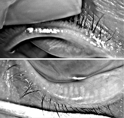
    I start questioning the accuracy of the images as I have strange discrepancy.

    My dr finally replied my email confirming the 1st images taken 10.2016 (by slit lamp camera by himself)
    the % area of loss (aol) is
    ''significantly higher'' than the 2nd images taken in 03.2017.
    Does not make sense, since glands wont grow, right?

    3rd images (05.2017) by infrared light % aol about 55%, exactly the same as 2nd images

    4th images (09.2017) by infrared light (same machine as the 3rd's)
    % alo (dropped to) about 28% (= more glands alive??)


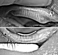
    -All are only lower glands. 3rd & 4th are from different doctors, 1st & 2nd are the same doctor
    -Did LipiFlow 07.2016 - felt great each day in 2-3 months but only lasted 3 weeks. All glands are open after LipiFlow, I was told. Unfortunately no images were taken prior to LipiFlow (although I paid for LipiView test).
    -Pain reduced about 70% after LipiFlow and no more pain for 7 months

    Seems the results also depend on HOW to roll the lids, which probably explain the discrepancy??
    Anyone has similiar experience? Any thoughts on this? My family said it is impossible to get 100% accuracy?
    Hard to tell from pictures as some are pretty small. I guess maybe 40% difference is easier to spot.

    Thanks!
    Last edited by MGD1701; 08-Jan-2018, 04:23.

  • #2
    Gland Recovery??
    I have found related report/video/images (dated Feb. 2015 & 2016), from Dr Arthur B. Epstein, OD, FAAO
    Any comments? Anyone has similiar experience or has read some reports on this?

    --------------------------------
    ''Off the Cuff: The Point of No Return
    ......
    Today, most experts believe that if left untreated, obstructed meibomian glands will down-regulate, cease functioning and ultimately wither and die. But when does a non-functioning meibomian gland become truly dead? That question has been a point of debate. Some believe that once non-functional, the meibomian gland is gone forever, while others — myself included — believe that
    glands can recover if treated before they reach the point of no return.

    Unblocking the meibomian glands and restoring function is not as simple as it might seem. Manual expression, even with heat, is marginally effective and only for some patients. Although proper management is a complex process, as many of you know, TearScience's LipiFlow system has become a core part of my therapeutic strategy. While I gauge success by patient response, I've found meibography to be extremely useful in assessing the severity of the disease and, as you will see in the following images, perhaps in the success of the treatment.

    Both images were captured using an Oculus Keratograph 5M. The first was pretreatment, the second taken about four months post LipiFlow. While the images are admittedly not perfect, the apparent increase in normal-looking meibomian glands, especially in the upper lid, is remarkable. The improvement in patient symptoms was likewise impressive.

    While this should not be taken as definitive evidence for recovery of “lost” meibomian glands after treatment, the images are compelling. We are currently following dozens of patients some with similar results.''
    https://www.reviewofoptometry.com/ne...ebruary-9-2015

    Interesting (educational) video (April 26. 2016) from the same dr, including:
    21:40 his treatment plans
    22:59 OTC drops he uses, including Systance Balance/Ultra
    https://www.youtube.com/watch?v=McVzkzOg5A4&t=21s
    Last edited by MGD1701; 11-Nov-2017, 05:43.

    Comment


    • #3
      I'm sorry I can't help with your query - but just wanted to thank you for the really helpful post above. I'm going to print-this out to take with me whenever I see opthalmologists. I was sent away for too long with the "just use hot compresses" advice, and I'm sure not being given better treatment is the reason why my glands/mgd/dry eye has got so bad.

      It's also hopeful to see that possibly glands might recover.

      Comment


      • #4
        + + + + + +
        Last edited by MGD1701; 05-Nov-2017, 13:36.

        Comment


        • #5
          Hey
          Visited a dr in a well-known University Eye Hospital who confirmed gland recovery is possible. Doctors there never took images nor osmolarity test (although they have them) so no idea if gland recovery is my case. He said my conditions are better than 3 months ago. I asked if LipiFlow, manual expression, IPL or debridement would help me. He said I do NOT need them - just continue with compress/massage 2 times/week (instead of 2/day) + drop, Retaine MGD. Meanwhile, I have never done probing as I do not have scar tissue. Doctors never prescribed me medication such as Ciclosporin (Ikervis), restasis etc - nor recommended Omega 3.

          My eyes have been pretty fit for 3 months, the best record, since I have tried:
          1) the lid cleanser, with pure Hypochlorous Acid, like Avenova or Heyedrate Lid & Lash (both only available in USA). For 1.5 months only needed 0-2 drops. Maybe you could also find alternatives - I have discovered 3 brands only the last one, NatraSan/made in UK, helps. Many/more American doctors recommend it so I was soso curious - very fascinating topic! What special is such pure HOCL has no resistance & effective/safe, according to doctors/Avenova.

          2) combination of Omega 3 +6/GLA (is more effective than omega 3 alone)
          Have read many articles from the American professional journals on this.
          For about 2 months I have noticed that there is more oil in the tear film, even when wake-up (before: hardly).
          However, my omega 3 & 6 levels are very high already, according to blood tests I initiated 1 year ago
          (Nevertheless, I have increased (bio, cold pressed) flaxseed oil 3 months ago - to thin the oil - instead of doxy which dr prescribed me 3 months ago for the first time but it is only as a last solution.

          We dont have InflammaDry, Sjo Tests nor Mibo. Manual expression is almost impossible. Osmolarity test, IPL, BlephEx and even doctors are hard to find. To get an appointment in hospitals 3-6 months waiting time. Most doctors only spend about 5 minutes and do not identify causes so what can I expect?? I have visited nearly 15 doctors and only 6 doctors know about MGD. Dont even know upper gland status. What sad is, only one doctor was honest telling me, ''Sorry, I can not help you.'', 2nd visit though - last year. I do appreciate her honesty because I finally realized I can NOT trust nor count on doctors anymore and have started my own research/experiments (literally day and night - but kind of fun to learn new things!).

          So, never give up - there are always solutions, if we search for them in a proactive and tenacious fashion!
          Not easy but possible!
          Last edited by MGD1701; 09-Apr-2018, 00:18.

          Comment


          • #6
            Hi,
            I've found that interesting:

            http://eyedoc2020.blogspot.fr/2016/1...meibomian.html
            http://eyedoc2020.blogspot.fr/2017/0...ibomian_5.html


            Comment


            • #7
              Thanks for sharing. Wondering WHY most doctors say the oppositive?
              It was pitty two of my doctors did not provide right/sufficient info at the time when I visited them, otherwise I should have known my status NOW. One even did not see the images - just read % the technican wrote - and it turned out what he told me was the oppositive.
              Last edited by MGD1701; 09-Apr-2018, 00:19.

              Comment


              • #8
                I know this feeling, I visited 7 doctors for 3 years before finding the good one who diagnosed my blepharis posterior (MGD) , I would not have lost 25% of my glands !
                Doctor said the glands will not come back, maybe because it is a very long process and they have to follow patients and make statisctics.
                I think patients like us should have a meibography (lipiview) every year and compare if some glands grows back.


                Comment


                • #9
                  Regarding the link you shared, that (American) dr mentioned that was her ONLY case (= only ONE) so far, right?
                  Dr Epstein mentioned in his video above that he had a number of gland recovery.
                  Well, at least now we know it is really possible!!
                  Last edited by MGD1701; 04-Nov-2017, 04:44.

                  Comment


                  • #10
                    Hey, update, I just received

                    1) formal report from the hospital indicating, both eyes show significant improvements & no inflammation (as usual) etc.
                    (also mentioned I should monitor my status in local clinics and only visit them when needed)

                    2) confirmation from the machine agent that their image print-outs ''only'' show ''area of loss''.
                    Why it is important? Because the dr (for the 4th images) did not see the images (nor showed me), just read % from pc. What worse is he said it was area of ALIVE but the print-out (sent by post) indicates: area of LOSS - huge difference - drove me nuts.

                    Last edited by MGD1701; 09-Apr-2018, 00:20.

                    Comment


                    • #11
                      Hi MGD1707,

                      Image:
                      Have you got all meibography images?
                      If you did a Lipiview the doctor can email you the image.
                      After receiving it you can by yourself estimate the area of loss.
                      Normally Lipiview should be done before making Lipiflow.
                      What can you see of the image ?

                      Lipiview can shows upper and lower eyelid meibographie + LLT + partial blinking %.
                      I think that if everybody could have a lipiview once a year we could follow the evolution (grow back) of MGs.

                      Doxy:
                      My doc gave me doxy for 6 weeks. Yes Doxy helps to thin the oil.

                      Hypochlorous Acid effect:
                      Reduction in bacterial load using hypochlorous acid hygiene solution on ocular skin
                      https://www.ncbi.nlm.nih.gov/pmc/articles/PMC5402722/


                      Sandra Lora Cremers Blog
                      http://eyedoc2020.blogspot.fr/
                      Another Patient who felt significant improvement after Lipiflow and meibography shows improvement
                      http://eyedoc2020.blogspot.fr/#!/201...gnificant.html

                      So regarding the MG grow back it is not the only one. Good hope


                      Comment


                      • #12
                        Hi
                        Images: I did LipiView prior to LipiFlow but dr did not show/tell me images. I asked if gland atrophy, dr said nothing. For me - just wasted money.

                        The problem is I dont know % of the 1st image as still waiting dr reply to my email which I sent after receipt of his recent confirmation that % area of LOSS was significantly higher (than 2nd's) - no idea when he will reply though. Sizes of 4th photos were too small to compare - I assume if difference is more than 35% then it is easier to spot.

                        To be honest, I am not that crazy about gland recovery as I still believe the accuracy also depends on HOW to ROLL the lids. However, the dr took the 1st & 2nd images is an well-known expert on such issue - but my family consider the 4th image should be more accurate. I will ask dr next time when a chance presents itself. What important are: my eyes are now manageable & I now finally know gland recovery is possible. Sometimes if we dont expect too much, maybe better outcomes would be possible??

                        Maybe others have similar cases to share???
                        Last edited by MGD1701; 09-Nov-2017, 12:07.

                        Comment


                        • #13
                          I was able to get some pictures taken for free via a Keratograph 5M machine at a local medical research university. Found it very helpful, not only the images of the meibomian glands but also the high detail of the imagery of the eye surface and tear film. Using the machine they did examinations of tear break up time, meniscus tear level, lipid layer, lissamine green and fluorescein dye stain tests. Staining showed I have scar folds on the conjunctiva that is dictating the placement of tears, and the usual punctate keratitis on the cornea, worse in my left eye. The meibomian glands only 3 partial glands could be picked up, though they said it's possible they can't pick up some very very tiny meibomian lines as they could see some signs of stuff at orifices that showed no identifiable duct line. They seemed to think I have some reflex tear production still perhaps, mainly in the right eye judging by meniscus level.

                          I had searched online for anywhere near me who had this machine and found some article about the university obtaining one and name of one of the researchers utilizing it who I then contacted over email. Worth seeing if there's anywhere who offers it, some public optician clinics do. I wonder if those who invest in a machine that has purely diagnostic purposes, compared to one that is typically used to pre-screen people for Lipiflow, may be more likely to offer more time / care in interpreting results but that may just be me being too cynical. Lipiview tests are certainly more available widely and even offered for free at some mainstream opticians.
                          Sufferer due to Toxic Epidermal Necrolysis.
                          Avatar art by corsariomarcio

                          Comment


                          • #14
                            Hi PhoenixEyes
                            Thanks for the interesting inputs. You are indeed lucky not only free images but also great diagnosis.
                            All doctors took images made no comments - only valuable info is % area of loss.

                            I have heard about Keratograph 5M - some doctors like it. The images above also from this machine.. Maybe I should find one next time. Many doctors in USA now use LipiScan (portable) - no ideas about difference though.
                            Last edited by MGD1701; 09-Nov-2017, 14:15.

                            Comment


                            • #15
                              hi mbperso
                              How interesting to know both of us have been prescribed doxy - and our doctors do not recommend omega 3.

                              I have read that 50mg x2/day is more effetive than 100mg/day, for your information.

                              Comment

                              Working...
                              X